MF3D Database
CC License
All MF3D data and media files are made available under the Creative Commons (CC) BY-NC-SA license. This license enables users to distribute, remix, adapt, and build upon the material in any medium or format for noncommercial purposes only, and only so long as attribution is given to the creator. If you remix, adapt, or build upon the material, you must license the modified material under identical terms. CC BY-NC-SA includes the following elements:
BY: Credit must be given to the creator.
NC: Only noncommercial uses of the work are permitted.
SA: Adaptations must be shared under the same terms.
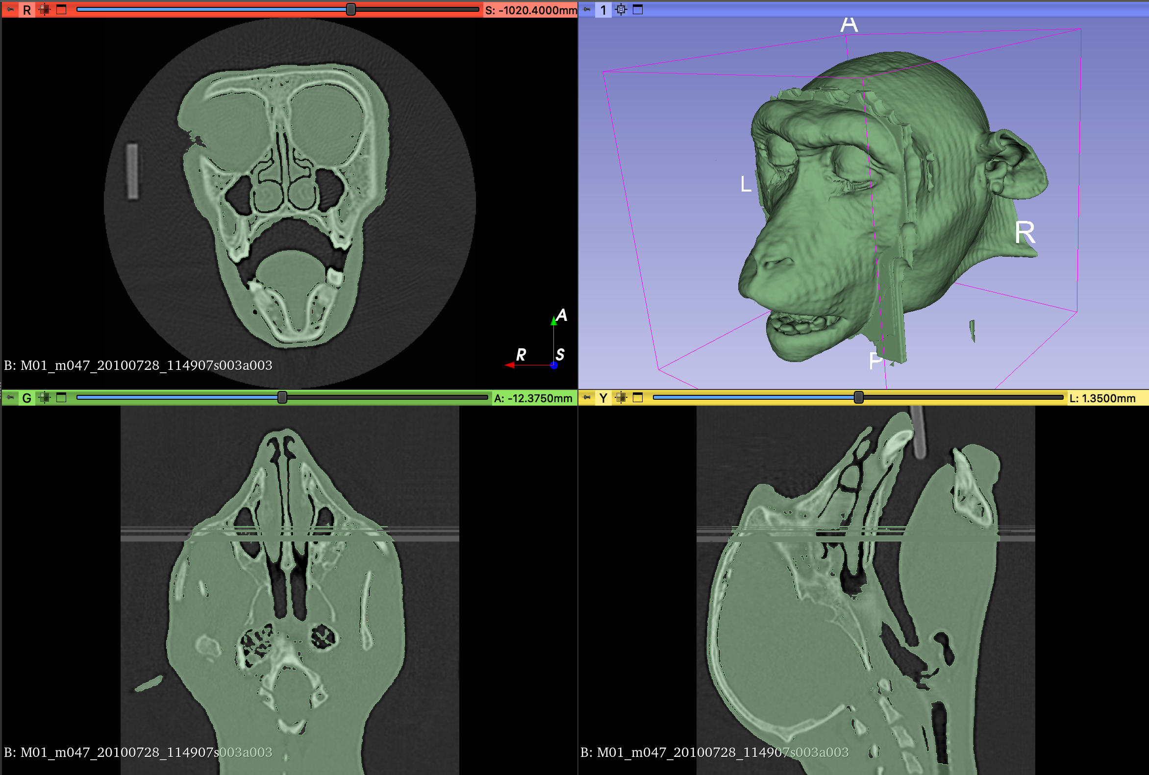
CT data acquisition

All 3D surface data used in MF3D databases were derived from in vivo computed tomography (CT) scans of anesthetized captive Rhesus macaque (m.mulatta) monkeys. Data were acquired at a range of research institutions, under protocols approved by local Institutional Animal Care and Use Committee (IACUC)’s that conform to national animal welfare law (e.g. the Animal Welfare Act in the US).
Database demographics
The validity of a morphable model for a given application is fundamentally limited by the sample data that were used to construct it. In particular, while the central tendency of a sample will quickly converge towards an accurate representation of the population mean, the accuracy of linearly interpolated morphs into the extreme periphery of the model space will suffer if that region is under sampled. For example, changes in craniofacial morphology with age are non-linear: there is an early period of bone and muscle growth, followed by loosening of the skin and eventual loss of muscle tone. The demographic make-up of the samples used to construct the original MF3D face-space and other morphable models are provided below.

The demographic make-up of the sample used to construct the original MF3D face-space is shown in the plot to the right. All animals in this sample (N = 36) were over 4 years of age, and only a small proportion were female (N = 7). The voxel resolution of CT volumes varied from 0.25 - 0.37mm in-plane, and 0.125 - 0.5mm slice thickness.


MF3D Demographics Table
ID |
Species |
Gender |
Weight (kg) |
Scan date |
DOB |
Age (yrs) |
Resolution in plane(mm) |
Slice thickness (mm) |
Source |
Source ID# |
Position |
Origin ID |
MRI |
Notes |
|---|---|---|---|---|---|---|---|---|---|---|---|---|---|---|
M01 |
Mulatta |
Female |
6.7 |
6 |
0.225 |
0.2 |
DMM |
47 |
Stereotax |
m047 |
||||
M02 |
Mulatta |
Male |
7.1 |
6 |
0.25 |
0.2 |
DMM |
48 |
Stereotax |
m048 |
||||
M03 |
Mulatta |
Male |
5.65 |
2/4/15 |
4/29/08 |
6.8 |
0.3 |
0.5 |
NIH |
FB81 |
Stereotax |
MrBeckey |
1 |
Headpost |
M04 |
Mulatta |
Male |
7.5 |
2/4/15 |
5/5/07 |
7.7 |
0.3 |
0.5 |
NIH |
DP9T |
Stereotax |
Pauling |
1 |
Headpost |
M05 |
Mulatta |
Male |
9.45 |
3/14/13 |
9 |
0.26 |
0.3 |
NIH |
DBKL |
Stereotax |
Alpha |
ECoG |
||
M06 |
Mulatta |
Female |
9.5 |
8/14/14 |
6 |
0.29 |
0.5 |
NIH |
KM2 |
Stereotax |
Snookie |
ECoG |
||
M07 |
Mulatta |
Female |
4.8 |
3/18/15 |
6 |
0.285 |
0.5 |
NIH |
K91 |
Stereotax |
Theta |
ECoG |
||
M08 |
Mulatta |
Male |
9.5 |
10/30/14 |
8 |
0.324 |
0.5 |
NIH |
DV74 |
Stereotax |
Zeta |
ECoG |
||
M09 |
Mulatta |
Male |
12 |
2/1/17 |
4/9/10 |
6.8 |
0.37 |
0.3 |
NIH |
DX8N |
Stereotax |
Hendrix |
1 |
Headpost |
M10 |
Mulatta |
Male |
7.7 |
9/15/16 |
4/7/08 |
8.4 |
0.35 |
0.5 |
NIH |
DV64 |
Stereotax |
Gumby |
Scar |
|
M11 |
Mulatta |
Female |
5.1 |
10/29/15 |
8/6/08 |
7.2 |
0.36 |
0.125 |
NIH |
KFZ |
Stereotax |
Regina |
1 |
Headpost |
M12 |
Mulatta |
Male |
12.5 |
12/15/16 |
6/2/09 |
7.5 |
0.37 |
0.25 |
NIH |
FC7P |
Stereotax |
Bennie |
Headpost |
|
M13 |
Mulatta |
Male |
10.1 |
6/27/17 |
4/3/12 |
5.2 |
0.37 |
0.25 |
NIH |
H818 |
Stereotax |
Jasper |
1 |
|
M14 |
Mulatta |
Male |
13.95 |
11/4/16 |
1/1/06 |
10.8 |
0.37 |
0.25 |
NIH |
DN4R |
Stereotax |
Agnew |
||
M15 |
Mulatta |
Male |
11.25 |
8/4/16 |
5/10/10 |
6.2 |
0.37 |
0.25 |
NIH |
DX7F |
Stereotax |
Bruce |
Headpost |
|
M16 |
Mulatta |
Male |
11.45 |
8/4/16 |
5/17/10 |
6.2 |
0.37 |
0.25 |
NIH |
GB7X |
Stereotax |
Christian |
Headpost |
|
M17 |
Mulatta |
Male |
7.6 |
8/27/15 |
6/7/10 |
5.2 |
0.293 |
0.5 |
NIH |
DX8L |
Stereotax |
Dobbs |
1 |
|
M18 |
Mulatta |
Male |
6 |
9/11/15 |
5/12/10 |
5.3 |
0.355 |
0.3 |
NIH |
DW7WX |
Stereotax |
Micro |
1 |
Headpost |
M19 |
Mulatta |
Male |
14.1 |
4/21/16 |
5/7/10 |
6 |
0.37 |
0.125 |
NIH |
DX4X |
Stereotax |
Piroette |
1 |
Headpost |
M20 |
Mulatta |
Male |
6.7 |
2/18/16 |
5/4/11 |
4.8 |
0.37 |
0.125 |
NIH |
H114 |
Stereotax |
Voltaire |
Headpost |
|
M21 |
Mulatta |
Male |
5.7 |
8/20/15 |
8/20/11 |
4 |
0.352 |
0.25 |
NIH |
H502 |
Stereotax |
Waldo |
Headpost |
|
M22 |
Mulatta |
Male |
7.3 |
5/10/18 |
4/9/14 |
4.1 |
NIH |
H47F |
Stereotax |
Wasabi |
1 |
Headpost |
||
M23 |
Mulatta |
Male |
8.5 |
12/7/18 |
7/17/12 |
6.4 |
0.37 |
0.25 |
NIH |
H753 |
Stereotax |
Zoboomafoo |
Headpost |
|
M24 |
Mulatta |
Male |
6.8 |
11/20/17 |
8/10/09 |
8.3 |
0.37 |
0.25 |
NIH |
FE5F |
Stereotax |
Prima |
1 |
Headpost |
M25 |
Mulatta |
Male |
8.5 |
12/28/14 |
5/22/09 |
5.6 |
0.293 |
0.25 |
NIH |
FC7V |
Stereotax |
Mega |
Headpost |
|
M26 |
Mulatta |
Male |
7.8 |
1/18/18 |
5/2/13 |
4.7 |
0.37 |
0.25 |
NIH |
H29L |
Stereotax |
Blis |
1 |
Headpost |
M27 |
Mulatta |
Male |
12.2 |
2/13/17 |
4/1/92 |
24.9 |
0.46 |
0.625 |
UC Davis |
MMU26667 |
Stereotax |
A |
||
M28 |
Mulatta |
Male |
14.2 |
3/15/17 |
6/1/91 |
25.8 |
0.31 |
0.625 |
UC Davis |
MMU26412 |
Stereotax |
B |
||
M29 |
Mulatta |
Male |
11.5 |
2/15/17 |
2/1/98 |
19 |
0.48 |
0.625 |
UC Davis |
MMU30529 |
Stereotax |
C |
||
M30 |
Mulatta |
Male |
13.1 |
4/21/14 |
3/1/00 |
14.1 |
0.4 |
0.625 |
UC Davis |
MMU32261 |
Stereotax |
D |
Headpost |
|
M31 |
Mulatta |
Male |
11.1 |
7/12/17 |
3/1/01 |
16.4 |
0.36 |
0.625 |
UC Davis |
MMU32891 |
Stereotax |
E |
Headpost |
|
M32 |
Mulatta |
10/15/14 |
0.34 |
0.625 |
UC Davis |
MMU21835 |
Stereotax |
F |
||||||
M33 |
Mulatta |
Male |
16 |
11/15/17 |
5/1/09 |
8.5 |
0.39 |
0.625 |
UC Davis |
MMU40100 |
Stereotax |
G |
Lips cropped |
|
M34 |
Mulatta |
Female |
7.5 |
10/9/14 |
2/1/05 |
9.7 |
0.34 |
0.625 |
UC Davis |
MMU36179 |
Stereotax |
H |
||
M35 |
Mulatta |
Female |
9.3 |
5/6/14 |
3/1/07 |
7.2 |
0.32 |
0.625 |
UC Davis |
MMU37884 |
Stereotax |
I |
||
M36 |
Mulatta |
Female |
7.8 |
10/8/13 |
3/1/03 |
10.6 |
0.29 |
0.625 |
UC Davis |
MMU34748 |
Stereotax |
J |
||

To extend the face-space model to more accurately cover the craniofacial morphology of infant macaques, we used data from the UNC-Wisconsin Rhesus macaque Neurodevelopment Database (Young et al., 2017). This database includes anatomical (T1-weighted) MRI scans from 36 infant Rhesus macaques between the ages of 2 weeks to 4 years old, collected longitudinally (150 scans total). The demographic distribution of this sample is shown in the plot on the right.
MF3Di Demographics Table
scan ID |
Sex |
DOB |
Birth weight (g) |
Matriline |
Patriline |
Age 1/days |
Scan 1 |
weight kg |
Age 2/days |
Scan 2 |
weight |
Age 3/days |
Scan 3 |
weight |
Age 2/days |
Scan 4 |
weight |
Age 2/days |
Scan 5 |
weight |
|---|---|---|---|---|---|---|---|---|---|---|---|---|---|---|---|---|---|---|---|---|
1 |
F |
6/6/10 |
438 |
1 |
1 |
360 |
06/01/11 |
1.9 |
493 |
10/12/11 |
2.6 |
605 |
02/01/12 |
2.76 |
758 |
07/03/12 |
3.16 |
863 |
10/16/12 |
3.4 |
2 |
M |
6/3/10 |
590 |
2 |
2 |
363 |
06/01/11 |
2.22 |
496 |
10/12/11 |
2.66 |
608 |
02/01/12 |
2.8 |
761 |
07/03/12 |
3.36 |
866 |
10/16/12 |
3.78 |
4 |
M |
3/11/10 |
590 |
4 |
4 |
482 |
07/06/11 |
3.08 |
601 |
11/02/11 |
3.26 |
727 |
03/07/12 |
4.02 |
853 |
07/11/12 |
4 |
972 |
11/07/12 |
4.94 |
5 |
F |
12/10/09 |
452 |
5 |
5 / 4 |
601 |
08/03/11 |
2.68 |
727 |
12/07/11 |
3.06 |
846 |
04/04/12 |
3.34 |
958 |
07/25/12 |
3.78 |
1090 |
12/04/12 |
4.66 |
6 |
M |
12/28/09 |
648 |
6 |
6 |
584 |
08/04/11 |
3.12 |
709 |
12/07/11 |
3.62 |
828 |
04/04/12 |
4.22 |
940 |
07/25/12 |
4.98 |
1072 |
12/04/12 |
5.47 |
7 |
M |
12/20/10 |
495 |
7 |
7 |
261 |
09/07/11 |
1.86 |
380 |
01/04/12 |
2.12 |
499 |
05/02/12 |
2.38 |
625 |
09/05/12 |
2.88 |
758 |
01/16/13 |
3.19 |
8 |
F |
12/29/10 |
460 |
8 |
8 |
252 |
09/07/11 |
1.86 |
371 |
01/04/12 |
2.1 |
490 |
05/02/12 |
2.6 |
616 |
09/05/12 |
3.28 |
750 |
01/17/13 |
3.81 |
10 |
M |
10/11/11 |
626 |
10 |
10 / 9 |
197 |
04/25/12 |
1.53 |
316 |
08/22/12 |
2.31 |
427 |
12/11/12 |
2.65 |
540 |
04/03/13 |
3.42 |
652 |
07/24/13 |
3.84 |
11 |
F |
12/15/10 |
425 |
11 |
4 |
498 |
04/26/12 |
2.32 |
616 |
08/22/12 |
2.82 |
721 |
12/05/12 |
3.17 |
847 |
04/10/13 |
3.48 |
973 |
08/14/13 |
4.12 |
12 |
F |
10/19/11 |
580 |
12 |
12 |
195 |
05/01/12 |
1.49 |
315 |
08/29/12 |
2.27 |
419 |
12/11/12 |
2.66 |
532 |
04/03/13 |
3.24 |
665 |
08/14/13 |
3.68 |
13 |
M |
12/13/11 |
616 |
13 |
13 |
148 |
05/09/12 |
1.2 |
266 |
09/04/12 |
2.12 |
407 |
01/23/13 |
2.94 |
505 |
05/01/13 |
3.13 |
617 |
08/21/13 |
3.56 |
14 |
F |
12/29/11 |
463 |
14 |
14 |
146 |
05/23/12 |
0.99 |
250 |
09/04/12 |
1.6 |
391 |
01/23/13 |
2.32 |
489 |
05/01/13 |
2.48 |
615 |
09/04/13 |
2.78 |
15 |
F |
2/2/12 |
420 |
15 |
14 / 9 |
132 |
06/13/12 |
1 |
||||||||||||
16 |
M |
2/23/12 |
672 |
16 |
1 |
118 |
06/20/12 |
1.26 |
244 |
10/24/12 |
1.88 |
349 |
02/06/13 |
2.16 |
475 |
06/12/13 |
2.56 |
606 |
10/21/13 |
2.98 |
17 |
M |
4/14/12 |
460 |
17 |
17 / 3 |
95 |
07/18/12 |
0.71 |
228 |
11/28/12 |
1.42 |
326 |
03/06/13 |
2 |
445 |
07/03/13 |
2.46 |
578 |
11/13/13 |
3 |
18 |
F |
4/27/12 |
609 |
18 |
44 |
103 |
08/08/12 |
1.01 |
229 |
12/12/12 |
1.52 |
313 |
03/06/13 |
2.04 |
432 |
07/03/13 |
2.34 |
566 |
11/14/13 |
3.06 |
19 |
F |
6/4/12 |
496 |
19 |
1 |
121 |
10/03/12 |
1.11 |
254 |
02/13/13 |
1.75 |
373 |
06/12/13 |
2.3 |
492 |
10/09/13 |
2.7 |
597 |
01/22/14 |
3.24 |
20 |
M |
9/23/12 |
480 |
20 |
20 |
64 |
11/26/12 |
0.89 |
170 |
03/12/13 |
1.34 |
233 |
05/14/13 |
1.62 |
332 |
08/21/13 |
2.24 |
423 |
11/20/13 |
2.35 |
21 |
F |
11/11/12 |
500 |
21 |
21 |
74 |
01/24/13 |
0.8 |
157 |
04/17/13 |
1.19 |
255 |
07/24/13 |
1.56 |
339 |
10/16/13 |
1.91 |
451 |
02/05/14 |
1.9 |
22 |
M |
12/26/12 |
570 |
22 |
9 / 4 |
57 |
02/21/13 |
0.74 |
134 |
05/09/13 |
1.28 |
273 |
09/25/13 |
2.08 |
356 |
12/17/13 |
2.12 |
434 |
03/05/14 |
2.62 |
23 |
F |
1/4/13 |
488 |
23 |
23 |
54 |
02/27/13 |
0.72 |
153 |
06/06/13 |
1.15 |
236 |
08/28/13 |
1.64 |
320 |
11/20/13 |
1.85 |
432 |
03/12/14 |
2.24 |
24 |
F |
2/22/13 |
510 |
24 |
24 |
26 |
03/20/13 |
0.56 |
||||||||||||
25 |
M |
2/22/13 |
540 |
25 |
23 |
27 |
03/21/13 |
0.68 |
117 |
06/19/13 |
1.1 |
229 |
10/09/13 |
1.58 |
320 |
01/08/14 |
2.07 |
397 |
03/26/14 |
2.3 |
26 |
F |
12/10/12 |
26 |
26 |
87 |
03/07/13 |
1.01 |
212 |
07/10/13 |
1.55 |
333 |
11/08/13 |
2.18 |
450 |
03/05/14 |
2.8 |
590 |
07/23/14 |
3.11 |
|
27 |
M |
12/20/12 |
541 |
1 |
27 |
97 |
03/27/13 |
0.86 |
202 |
07/10/13 |
1.24 |
217 |
07/25/13 |
1.38 |
461 |
03/26/14 |
2.17 |
580 |
07/23/14 |
2.73 |
28 |
M |
3/12/13 |
582 |
28 |
9 |
29 |
04/10/13 |
0.71 |
127 |
07/17/13 |
1.24 |
218 |
10/16/13 |
1.6 |
309 |
01/15/14 |
2.12 |
400 |
04/16/14 |
2.74 |
29 |
M |
12/17/12 |
546 |
29 |
24 |
122 |
04/18/13 |
1 |
254 |
08/28/13 |
1.74 |
359 |
12/11/13 |
2 |
478 |
04/09/14 |
2.42 |
611 |
08/20/14 |
3 |
30 |
F |
1/4/13 |
516 |
30 |
30 |
116 |
04/30/13 |
1.1 |
250 |
09/11/13 |
1.66 |
376 |
01/15/14 |
2.31 |
488 |
05/07/14 |
2.79 |
600 |
08/27/14 |
3.18 |
31 |
M |
3/31/13 |
500 |
31 |
31 |
32 |
05/02/13 |
0.61 |
122 |
07/31/13 |
0.98 |
222 |
11/08/13 |
1.63 |
297 |
01/22/14 |
2.08 |
402 |
05/07/14 |
2.56 |
32 |
F |
5/13/13 |
492 |
32 |
32 |
17 |
05/30/13 |
0.54 |
102 |
08/23/13 |
0.96 |
198 |
11/27/13 |
1.54 |
289 |
02/26/14 |
2.03 |
373 |
05/21/14 |
2.34 |
33 |
F |
5/7/13 |
568 |
33 |
33 |
28 |
06/04/13 |
0.7 |
204 |
11/27/13 |
1.5 |
295 |
02/26/14 |
2.1 |
393 |
06/04/14 |
2.48 |
|||
34 |
M |
6/26/13 |
476 |
18 |
18 |
16 |
07/12/13 |
0.54 |
98 |
10/02/13 |
0.84 |
224 |
02/05/14 |
1.56 |
294 |
04/16/14 |
1.88 |
371 |
07/02/14 |
2.02 |
35 |
M |
7/23/13 |
622 |
35 |
35 |
13 |
08/05/13 |
0.69 |
93 |
10/24/13 |
1.13 |
204 |
02/12/14 |
1.66 |
281 |
04/30/14 |
2.03 |
349 |
07/07/14 |
2.49 |
36 |
M |
7/26/13 |
565 |
36 |
36 |
18 |
08/13/13 |
0.64 |
96 |
10/30/13 |
1 |
201 |
02/12/14 |
1.5 |
278 |
04/30/14 |
2.02 |
346 |
07/07/14 |
2.55 |

To extend the theoretical concept underlying ‘face-space’ to macaque bodies, we used a sample of whole-body CT data from 200 adult Rhesus macaques from the CNPRC, that was acquired by researchers at UC Davis (Buck et al., 2021). A subset of these data are available via Morphosource. All data were acquired on a GE Discovery® 610 PET/CT scanner, at an in-plane voxel resolution of 0.76mm and 0.625mm slice interval.
MB3D Demographics Table
Specimen |
Sex |
Yrs |
Mos |
kg |
|---|---|---|---|---|
29999 |
F |
18 |
11 |
9.3 |
30171 |
F |
20 |
0 |
8.49 |
30797 |
F |
17 |
11 |
12.6 |
31603 |
F |
16 |
8 |
7.35 |
31692 |
F |
16 |
11 |
7.7 |
32200 |
F |
16 |
9 |
8.5 |
32208 |
M |
16 |
0 |
14.8 |
32277 |
F |
15 |
8 |
7 |
32397 |
M |
16 |
11 |
14.85 |
32977 |
M |
15 |
0 |
17.3 |
33422 |
F |
14 |
6 |
8.87 |
33428 |
F |
14 |
6 |
11.44 |
33792 |
F |
14 |
10 |
10.8 |
33843 |
F |
14 |
0 |
9.46 |
34993 |
F |
12 |
5 |
10.09 |
35560 |
F |
11 |
11 |
10.75 |
35602 |
F |
11 |
11 |
5.93 |
35761 |
F |
11 |
0 |
10.93 |
35837 |
M |
11 |
9 |
11.53 |
35840 |
F |
11 |
6 |
9.45 |
36384 |
F |
11 |
0 |
6.6 |
36496 |
M |
10 |
10 |
16.02 |
36720 |
F |
10 |
10 |
9.42 |
36819 |
F |
11 |
8 |
10.87 |
37085 |
F |
9 |
11 |
11.15 |
37163 |
F |
10 |
3 |
12.58 |
37166 |
F |
10 |
3 |
8.42 |
37177 |
F |
9 |
0 |
9.53 |
37214 |
F |
10 |
3 |
9.9 |
37266 |
F |
9 |
11 |
9.66 |
37345 |
M |
9 |
10 |
13.26 |
37410 |
F |
9 |
3 |
7.12 |
37414 |
F |
9 |
7 |
6.77 |
37447 |
F |
12 |
1 |
11.78 |
37452 |
M |
9 |
10 |
18.6 |
37477 |
F |
9 |
7 |
5.3 |
37538 |
F |
9 |
10 |
11.34 |
37585 |
F |
9 |
9 |
7.57 |
37599 |
F |
9 |
9 |
5.53 |
37652 |
F |
10 |
1 |
15.83 |
37687 |
F |
10 |
0 |
7.38 |
37748 |
F |
10 |
0 |
8.9 |
37861 |
F |
9 |
3 |
7.2 |
37896 |
F |
9 |
3 |
9.49 |
38037 |
F |
8 |
11 |
10.83 |
38120 |
F |
9 |
10 |
7.43 |
38219 |
M |
9 |
11 |
14.95 |
38228 |
F |
8 |
10 |
5.8 |
38252 |
F |
8 |
10 |
11.18 |
38328 |
F |
8 |
10 |
15.21 |
38341 |
F |
8 |
10 |
7.66 |
38465 |
F |
9 |
0 |
9.5 |
38752 |
M |
8 |
2 |
11.84 |
38982 |
M |
7 |
7 |
8.96 |
39026 |
F |
7 |
11 |
7.26 |
39242 |
F |
7 |
9 |
7.39 |
39374 |
F |
7 |
9 |
7.91 |
39481 |
F |
7 |
6 |
9.41 |
39512 |
M |
7 |
8 |
9.58 |
39612 |
F |
7 |
0 |
7 |
39650 |
M |
7 |
2 |
11.45 |
39847 |
F |
6 |
11 |
7.36 |
39971 |
M |
6 |
10 |
11.9 |
40000 |
M |
6 |
10 |
11.21 |
40021 |
F |
6 |
10 |
7.33 |
40052 |
F |
7 |
0 |
11.23 |
40099 |
M |
7 |
1 |
11.69 |
40109 |
F |
6 |
10 |
6.39 |
40202 |
M |
6 |
9 |
10.88 |
40243 |
F |
6 |
9 |
5.18 |
40446 |
M |
6 |
7 |
10.93 |
40458 |
F |
6 |
4 |
5.84 |
41251 |
F |
6 |
2 |
6.4 |
41255 |
M |
7 |
1 |
9.6 |
28446 |
M |
23 |
3 |
11.51 |
31538 |
M |
18 |
1 |
10.57 |
33030 |
F |
17 |
1 |
9.71 |
33326 |
F |
16 |
3 |
7.14 |
33737 |
M |
15 |
2 |
15.47 |
33899 |
F |
16 |
1 |
9.34 |
33915 |
F |
15 |
3 |
8.36 |
34040 |
F |
15 |
0 |
9.28 |
34839 |
F |
15 |
0 |
9.73 |
34880 |
M |
15 |
0 |
12.61 |
34884 |
M |
15 |
0 |
16.58 |
35538 |
F |
13 |
0 |
8.51 |
35830 |
F |
13 |
3 |
8.42 |
36122 |
F |
13 |
2 |
8.44 |
36312 |
F |
12 |
3 |
9.75 |
36422 |
M |
13 |
0 |
19.76 |
37314 |
F |
12 |
1 |
9.44 |
37365 |
F |
11 |
3 |
7.9 |
37420 |
F |
11 |
3 |
9.62 |
37443 |
M |
11 |
3 |
12.01 |
37568 |
M |
11 |
3 |
9.03 |
37644 |
F |
10 |
3 |
5.24 |
37847 |
F |
11 |
1 |
11.19 |
37943 |
F |
10 |
3 |
8.29 |
38021 |
F |
11 |
0 |
14.82 |
38333 |
F |
10 |
3 |
8.5 |
38361 |
F |
11 |
0 |
6.94 |
38699 |
F |
10 |
1 |
12.65 |
38735 |
F |
10 |
1 |
7.81 |
39018 |
F |
9 |
1 |
7.21 |
39278 |
F |
9 |
3 |
9.61 |
39577 |
F |
9 |
1 |
12.99 |
39670 |
F |
9 |
1 |
5.35 |
39810 |
F |
9 |
1 |
7.32 |
39875 |
F |
8 |
3 |
5.39 |
39925 |
F |
9 |
0 |
12.04 |
39995 |
F |
9 |
1 |
9.05 |
40262 |
F |
8 |
3 |
12.14 |
40765 |
F |
7 |
3 |
7.86 |
40809 |
F |
8 |
1 |
8.38 |
41026 |
M |
7 |
3 |
9.88 |
41066 |
M |
7 |
3 |
12.26 |
41252 |
F |
7 |
3 |
7.86 |
42496 |
F |
6 |
1 |
5.62 |
42687 |
F |
6 |
0 |
5.14 |
42946 |
F |
5 |
3 |
8.05 |
44339 |
M |
4 |
0 |
7.86 |
44417 |
M |
4 |
0 |
7.4 |
45407 |
F |
2 |
3 |
6.02 |
46123 |
F |
5 |
3 |
8.2 |
46124 |
F |
5 |
1 |
6.65 |
46125 |
M |
5 |
3 |
9.74 |
46126 |
M |
5 |
1 |
11.25 |
46127 |
M |
7 |
0 |
11.88 |
46128 |
M |
6 |
1 |
13.72 |
46132 |
F |
6 |
1 |
5.4 |
46133 |
F |
6 |
1 |
6.3 |
46134 |
F |
6 |
1 |
6.4 |
46135 |
F |
6 |
1 |
7 |
46136 |
M |
6 |
1 |
12.57 |
46137 |
M |
6 |
1 |
12.96 |
46769 |
F |
17 |
0 |
11.07 |
32663 |
F |
18 |
4 |
9.94 |
32665 |
F |
18 |
4 |
10.27 |
32924 |
F |
17 |
7 |
11.33 |
33199 |
F |
17 |
6 |
7.37 |
33669 |
F |
16 |
8 |
13.46 |
46770 |
F |
17 |
6 |
12.1 |
37535 |
F |
12 |
5 |
12.95 |
33002 |
F |
17 |
7 |
11.34 |
32888 |
F |
17 |
7 |
12.36 |
27750 |
F |
25 |
4 |
9.62 |
33077 |
M |
17 |
6 |
11.63 |
32107 |
F |
18 |
7 |
10.79 |
32237 |
F |
18 |
7 |
8.55 |
32157 |
F |
18 |
7 |
10.21 |
32191 |
F |
18 |
7 |
6.61 |
32046 |
F |
18 |
8 |
8.98 |
32114 |
F |
18 |
7 |
8.95 |
32636 |
F |
18 |
4 |
7.98 |
29658 |
F |
22 |
5 |
7.23 |
34453 |
F |
24 |
2 |
8.35 |
32394 |
F |
18 |
7 |
11.1 |
31459 |
F |
19 |
8 |
9.04 |
34123 |
F |
21 |
5 |
7.11 |
37576 |
F |
12 |
7 |
9.95 |
41337 |
F |
8 |
5 |
8 |
35122 |
F |
15 |
7 |
10.28 |
41373 |
F |
8 |
4 |
9.12 |
39696 |
M |
9 |
9 |
19.2 |
34220 |
M |
16 |
7 |
17.11 |
33734 |
M |
16 |
9 |
14.91 |
37175 |
M |
12 |
8 |
17.62 |
37394 |
F |
12 |
7 |
6.22 |
37430 |
F |
12 |
8 |
11.18 |
37749 |
F |
12 |
5 |
13.34 |
43016 |
F |
6 |
6 |
6.33 |
37389 |
F |
12 |
8 |
11.88 |
42622 |
F |
6 |
8 |
6.07 |
41165 |
M |
8 |
7 |
10.76 |
31775 |
F |
19 |
9 |
9.51 |
31761 |
F |
19 |
9 |
6.83 |
31677 |
F |
19 |
10 |
10.31 |
31375 |
F |
19 |
11 |
7.37 |
30878 |
F |
20 |
10 |
11.64 |
39584 |
M |
10 |
3 |
13.52 |
40144 |
M |
10 |
1 |
14.47 |
39302 |
M |
11 |
1 |
14.5 |
39272 |
M |
11 |
1 |
20.4 |
40223 |
M |
10 |
1 |
22.35 |
42401 |
M |
7 |
2 |
16.7 |
42272 |
M |
7 |
4 |
10.81 |
42384 |
M |
7 |
2 |
11.85 |
40769 |
M |
9 |
2 |
20.97 |
37161 |
F |
13 |
4 |
11.09 |
39869 |
F |
10 |
3 |
7.64 |
40730 |
F |
9 |
4 |
10.17 |
39107 |
F |
11 |
3 |
11.35 |
37292 |
F |
13 |
3 |
13.36 |
38756 |
F |
11 |
4 |
5.84 |
38529 |
F |
12 |
1 |
11.33 |
36879 |
F |
14 |
1 |
12.36 |
Data Downloads
Request Data Access
Unlike the publicly available MF3D stimuli media files and MF3D code, all other MF3D data are are currently hosted on a password protected secure server. To request access to download MF3D data files, please contact New Atlantis Labs via the contact form below.
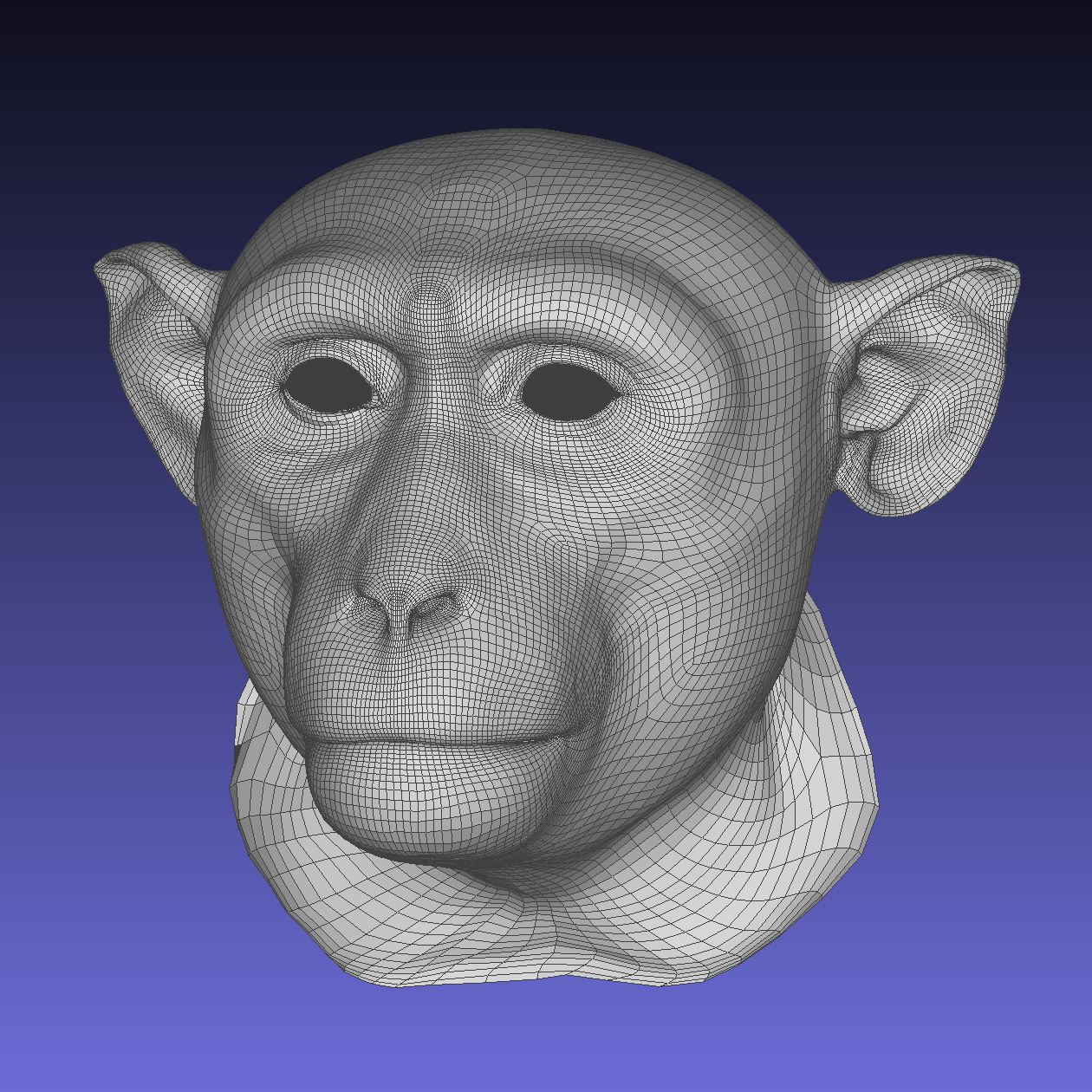
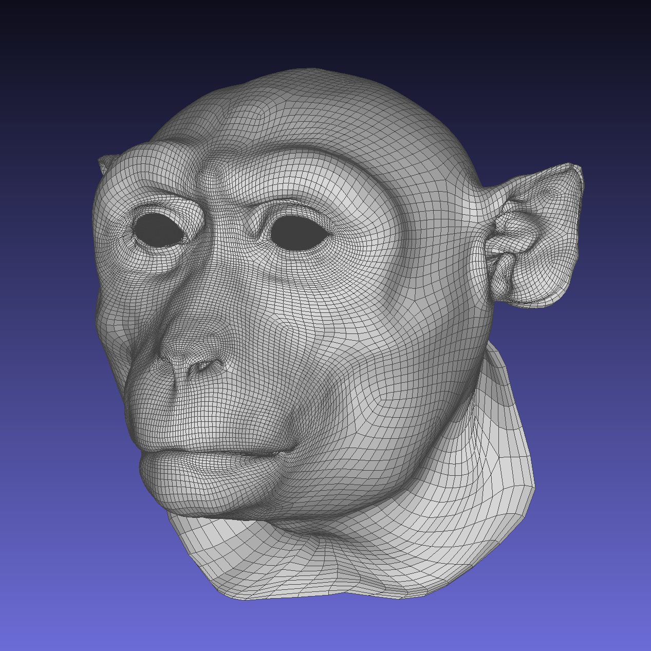
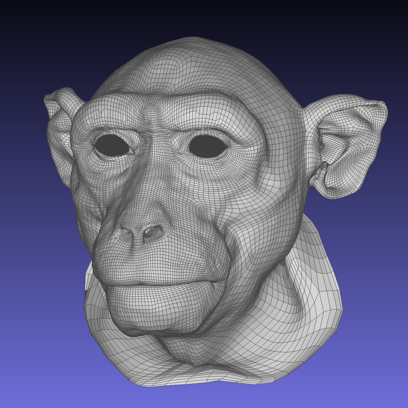
These standardized 3D surface meshes of macaque heads have identical uniform topology and polygon count (~50K), and are provided in .obj file format. They contain data about the 3D structure of each animal’s cranio-facial morphology, but no texture (e.g. color, fur, etc.) information. All faces have been edited to have open eye lids and neutral closed mouth expressions.
Average mesh
MF3D R1 ‘average’ identity in ‘neutral’ expression.
M01 mesh
MF3D identity ‘M01’ in ‘neutral’ expression.
M02 mesh
MF3D identity ‘M02’ in ‘neutral’ expression.